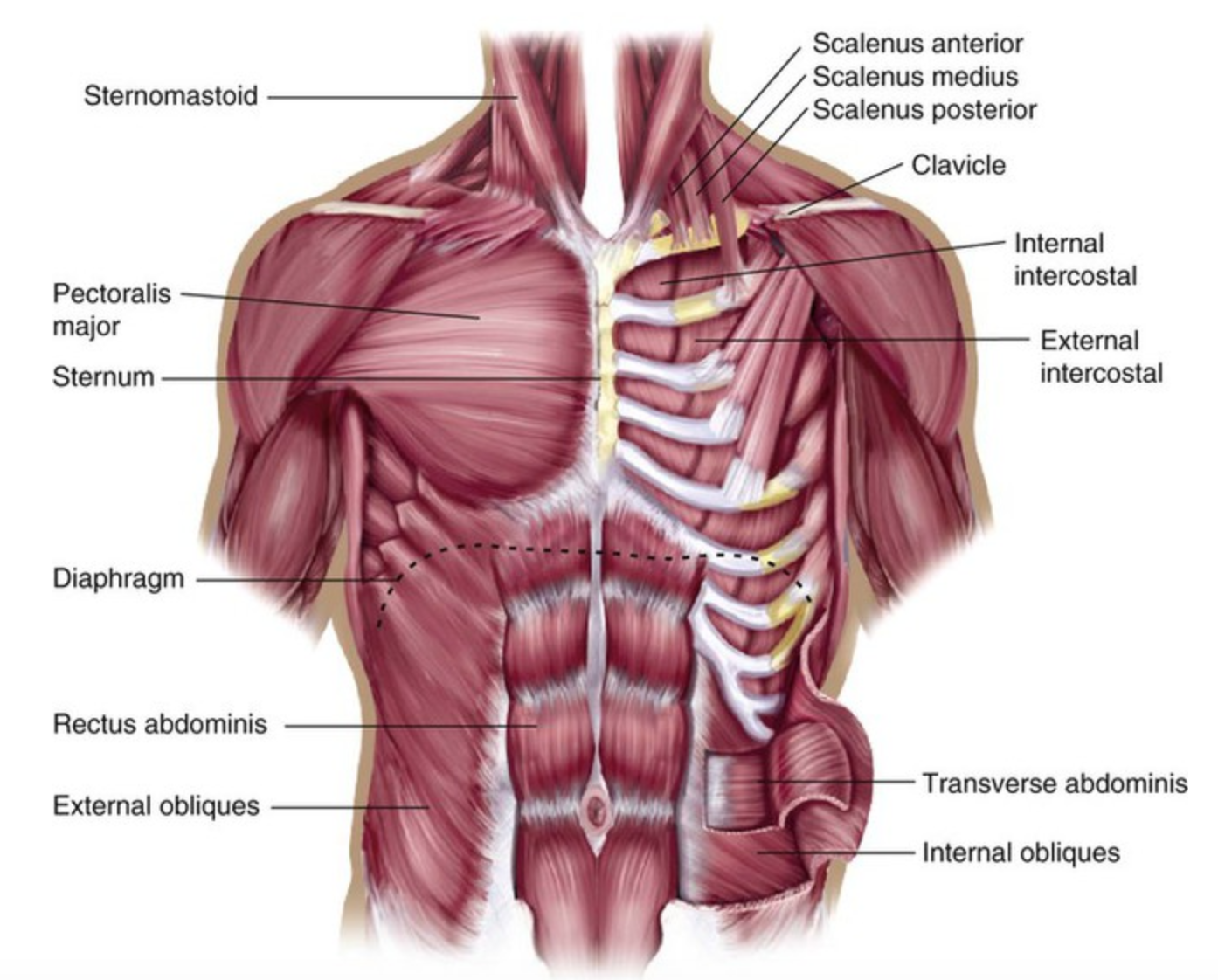
Diagram Of Chest Area : Human Chest Anatomy Images Stock Photos Vectors Shutterstock - Located high up at the tip of the cliff.. Diagram of a chest tube draining fluid from a plural effusion. The abdominal cavity is the part of the body that houses the stomach, liver, pancreas, kidneys, gallbladder, spleen, and the large and small intestines.the diaphragm marks the top of the abdomen and the horizontal line at the level of the top of the pelvis marks the bottom. Thoracic cavity, also called chest cavity, the second largest hollow space of the body.it is enclosed by the ribs, the vertebral column, and the sternum, or breastbone, and is separated from the abdominal cavity (the body's largest hollow space) by a muscular and membranous partition, the diaphragm.it contains the lungs, the middle and lower airways—the tracheobronchial tree—the heart. The major muscle in the chest is the pectoralis major. The anatomy of the human ribs is made up of 24 ribs which are parted in 12 pairs (each on the left and right side of the chest wall), with the sternum, metasternum(the.
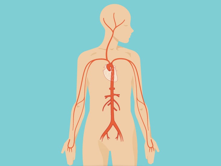
Your heart is surrounded by important blood vessels and arteries which pump blood into and out of your heart. A woman's chest — like the rest of her body — is covered with skin that has two layers. 12 cm (5 in) in length, 8 cm (3.5 in) wide, and 6 cm (2.5 in) in thickness. The nervous system of the thorax is a vital part of the nervous system as a whole, as it includes the spinal cord, peripheral nerves, and autonomic ganglia that communicate with and control many vital organs. Diagram of the emergence of the bronchial arteries in the descending thoracic aorta.
Female Chest Muscles Anatomy Diagram Function Body Maps from post.healthline.com The ribs and sternum make up what is called the 'ribcage.' the ribcage protects the lungs, blood vessels, and heart. The myofascial pain pattern has pain locations that are displayed in red and associated trigger points shown as xs. The diagrams depict the human eyeand light waves hitting the fovea, the area of detailed vision. Related posts of anatomy of the chest area acupressure points body picture. They make up the lateral part of our body, its anterior and posterior wall and they entirely build the lateral parts of the chest wall. Possible causes of pain include trauma, musculoskeletal. The anatomy of the human ribs (costae) are one of the integral parts of the chest wall; Chest pain doesn't always mean you're having a heart attack.
1 now that you know the exact place where the heart.
Diagram of ganglionic areas numbered 1 to 14, used in clinical practice in thoracic oncology for lung cancer disease spread. Strains and inflammation in the chest wall muscles or ribs usually goes away on its own. Possible causes of pain include trauma, musculoskeletal. The sternum, or breastbone, is a flat bone at the front center of the chest. Connective tissue called the mesentery holds the abdominal organs together. A typical heart is approximately the size of your fist: The circulatory system does most of its work inside the chest. Diagram of a chest tube draining fluid from a plural effusion. So for example, if the central angle was 90°, then the sector would have an area equal to one quarter of the whole circle. 12 cm (5 in) in length, 8 cm (3.5 in) wide, and 6 cm (2.5 in) in thickness. Diagram of chest area posted on july 2, 2015 by admin when we are doing cpr why do compress the sternum area not rh quora com human chest anatomy chest diagram of the chest area including lungs, heart (hidden by the lungs) and ribcage. Any diaphragm pain can, therefore, be very alarming. They make up the lateral part of our body, its anterior and posterior wall and they entirely build the lateral parts of the chest wall.
12 cm (5 in) in length, 8 cm (3.5 in) wide, and 6 cm (2.5 in) in thickness. Located high up at the tip of the cliff. The anatomy of the human ribs (costae) are one of the integral parts of the chest wall; Diagram of normal airway anatomy, frontal view. The ribs and sternum make up what is called the 'ribcage.' the ribcage protects the lungs, blood vessels, and heart.
Auckland Physiotherapy Limited Are You Breathing Wrong from lh6.googleusercontent.com And the other set of networks were. Diagram of chest area posted on july 2, 2015 by admin when we are doing cpr why do compress the sternum area not rh quora com human chest anatomy chest diagram of the chest area including lungs, heart (hidden by the lungs) and ribcage. So for example, if the central angle was 90°, then the sector would have an area equal to one quarter of the whole circle. The nervous system of the thorax is a vital part of the nervous system as a whole, as it includes the spinal cord, peripheral nerves, and autonomic ganglia that communicate with and control many vital organs. Check here to understand the function and part of it. A typical heart is approximately the size of your fist: The sternum, or breastbone, is a flat bone at the front center of the chest. Located high up at the tip of the cliff.
They make up the lateral part of our body, its anterior and posterior wall and they entirely build the lateral parts of the chest wall.
Acupressure points body picture 13 photos of the acupressure points body picture accupressure points the body, acupressure points body chart, acupressure pressure points, acupressure vs acupuncture, acupuncture point between thumb and index finger, how many acupuncture points on the body, message acupressure, human. Thoracic cavity, also called chest cavity, the second largest hollow space of the body.it is enclosed by the ribs, the vertebral column, and the sternum, or breastbone, and is separated from the abdominal cavity (the body's largest hollow space) by a muscular and membranous partition, the diaphragm.it contains the lungs, the middle and lower airways—the tracheobronchial tree—the heart. Shape and size of the heart. The nervous system of the thorax is a vital part of the nervous system as a whole, as it includes the spinal cord, peripheral nerves, and autonomic ganglia that communicate with and control many vital organs. Sensory information from the body and critical signals. The ribs and sternum make up what is called the 'ribcage.' the ribcage protects the lungs, blood vessels, and heart. The shape of the heart is similar to a pinecone, rather broad at the superior surface and tapering to the apex. The pectoralis major originates along the clavicle, down the sternum, and across the ribs and inserts into the humerus. Your heart is surrounded by important blood vessels and arteries which pump blood into and out of your heart. Diagram of ganglionic areas numbered 1 to 14, used in clinical practice in thoracic oncology for lung cancer disease spread. A woman's chest — like the rest of her body — is covered with skin that has two layers. In fact every radiologst should be an expert in chest film reading. Rectangular prisms, triangular prisms, trapezoidal prisms, hexagonal the following diagrams show a triangular prism and a rectangular prism.
Human chest bone structure parts of the chest bones. Thoracic cavity, also called chest cavity, the second largest hollow space of the body.it is enclosed by the ribs, the vertebral column, and the sternum, or breastbone, and is separated from the abdominal cavity (the body's largest hollow space) by a muscular and membranous partition, the diaphragm.it contains the lungs, the middle and lower airways—the tracheobronchial tree—the heart. A chest radiograph in isolation is limited in its contribution to diagnosis, and its usefulness is enhanced right neck paratracheal area aortic arch right hilum left hilum vascular shadows (aorta; Find out from webmd about other health problems that could be to blame. The abdominal cavity is the part of the body that houses the stomach, liver, pancreas, kidneys, gallbladder, spleen, and the large and small intestines.the diaphragm marks the top of the abdomen and the horizontal line at the level of the top of the pelvis marks the bottom.
Human Shoulder Anatomy Koibana Info Shoulder Muscle Anatomy Shoulder Anatomy Muscle Diagram from i.pinimg.com Diagram of normal airway anatomy, frontal view. The myofascial pain pattern has pain locations that are displayed in red and associated trigger points shown as xs. Human chest bone structure parts of the chest bones. Acupressure points body picture 13 photos of the acupressure points body picture accupressure points the body, acupressure points body chart, acupressure pressure points, acupressure vs acupuncture, acupuncture point between thumb and index finger, how many acupuncture points on the body, message acupressure, human. A woman's chest — like the rest of her body — is covered with skin that has two layers. Your heart is surrounded by important blood vessels and arteries which pump blood into and out of your heart. Sensory information from the body and critical signals. Related posts of anatomy of the chest area acupressure points body picture.
And the other set of networks were.
So for example, if the central angle was 90°, then the sector would have an area equal to one quarter of the whole circle. And the other set of networks were. A typical heart is approximately the size of your fist: Connective tissue called the mesentery holds the abdominal organs together. Chest pain doesn't always mean you're having a heart attack. A chest muscle that pulls the arm in towards the body. The epidermis is the outermost layer that provides a protective, waterproof seal over the body. The chest workout for huge, defined pecs. Understanding the basics of throat anatomy with diagram and pictures. For many, the chest is made up of a single rigid bone called the sternum.however, this is not true.other than the sternum, there are other bones in the chest region, such as the ribs and even the spine at the back. The anatomy of the human ribs (costae) are one of the integral parts of the chest wall; Possible causes of pain include trauma, musculoskeletal. The nervous system of the thorax is a vital part of the nervous system as a whole, as it includes the spinal cord, peripheral nerves, and autonomic ganglia that communicate with and control many vital organs.